Blog category: McDermott's Musings

Episode 6. Douchebag Department of the Week!


Another first for this series: this story is about a department – so, it’s about collective stupidity rather than just individual incompetence.

Yesterday I saw a lovely lady in her 60’s who’s got patellofemoral arthritis in her knee. Over the last 10 months she’s had 7 episodes where her knee has locked up, with severe pain. The longest episode of locking lasted 3 days.

This lady went to the orthopaedic department in her local hospital in West London. (This place is an utter ‘Pit of Doom’! (I know: many years ago I used to work there… and it was GRIM!)

  • On one occasion this lady’s knee has been manipulated in A&E, to unlock it. She was then simply given a knee brace and discharged.
  • On another occasion, the team that she saw in the orthopaedic department of her local hospital offered her a manipulation of her knee under a general anaesthetic (but without an arthroscopy at the same time!).
  • On one other occasion, she was offered an intra-articular injection of steroid.
  • She was also sent for a CT scan of her knee (quite why, I’m not sure)… but not an MRI!

Thankfully, the lady came to see me for a proper, sensible, sane opinion.

An MRI scan confirmed evidence of the pre-existing patellofemoral arthritis, and it showed that the medial and lateral menisci and the articular cartilage in the medial and lateral compartments all looked fine. However, there was clear evidence of a roughly 1cm-diameter bony loose body sitting at the front of the joint, directly in front of the ACL tibial footprint.

I’ve offered this lady a slot on my list next week, for an arthroscopy with removal of the loose body. (This should be a quick, easy, straightforward procedure, under a brief anaesthetic, done as a day-case, and she’ll be fully-weight-bearing straightaway afterwards.)

Learning points?

  1. If someone complains of recurrent temporary intermittent locking of their knee… then it’s a loose body or an unstable meniscal tear…. until proven otherwise!
  2. (If someone presents with a locked knee, especially after a twisting episode), then it’s a displaced locked bucket handle tear, until proven otherwise – and this is a far more urgent issue: https://kneearthroscopy.co.uk/condition/locked-knees/.)
  3. The correct imaging for this lady, right from the go, should have been an MRI scan.
  4. As a surgeon, if you’re going to give someone an anaesthetic, in theatre, to manipulate a locked knee straight… then why on Earth would you not also combine this, then and there, with a quick arthroscopy too, to remove the loose body (or, potentially, to deal with an unstable meniscal tear) at the same time, in order to fix the actual problem.
  5. Well done to A&E for giving this lady a brace…. as opposed to them doing absolutely nothing of any use or value whatsoever, which is so often the actual case for so many ‘orthopaedic’ patients (even if just giving her a brace was little more than just a token gesture!).
  6. Simply treating a knee with recurrent intermittent locking with an intra-articular steroid injection shows a fundamental and crushing lack of understanding about what the heck was actually going on in this poor lady’s knee.

Questions

  1. Would you like to know the name of the hospital in West London where this lady was mismanaged?
  2. Assuming that this lady does actually go ahead with the arthroscopy that I’ve offered her next week… then would you like to see a photo of the loose body?


Douchebag Doctor of the Week. Episode 5… also known this time as: ‘Awesome Physio of the Week!’


I’m ‘excited’ about this ‘Douchebag’ story – because this time we’ve got a special guest: the patient’s physio!

Luckily, this patient was fortunate enough to have a physio who’s senior enough, confident enough and decent enough to stand up against a very senior consultant by raising a red flag and encouraging the patient to get a second opinion.

Personally, I was shocked and appalled (again!) by what this patient told me. What do you think?….

Case History

History

An 86-year-old gentlemen was complaining of gradually increasing pain and stiffness in his thigh muscles, with increasing difficulty walking.

He went to see a Professor of Orthopaedics at an internationally famous and very flash clinic / hospital in Central London. The patient was listed for bilateral simultaneous knee replacement surgery.

As noted above, the patient’s physio was deeply concerned about this proposed plan of action, and hence he asked me to see the patient for a second opinion. At the time that I saw the patient in clinic he was, at that point, booked in to have this surgery just the following week.

When I called the patient’s name for him to come into my consulting room from the waiting area, it took the patient a while to get up from his seat, and it took him a while to get going… before he was then able to walk into my room reasonably well.

The patient said that he had pain in his knees that tended to come and go, but at worse, it was only “moderate” in severity. The patient felt this pain mainly on stairs, and he therefore tended to go up and down stairs just one step at a time. However, the patient was still able to go out for his usual ½-hour walk every day. Importantly, he stated that when he starts to walk he feels very stiff and he finds it difficult to get going; however, as he gets going his muscles then start to feel better, and that he then feels more mobile and a lot better. The patient was not having to take any painkillers or anti-inflammatories, and his knees were not affecting his sleep. The patient stated that he was not doing any other exercise at all apart from his usual ½-hour once-a-day walk.

Unfortunately, the patient had a long list of medical problems, including atrial fibrillation (for which he was on blood thinners), major bowel issues (with colitis, and with him having had a colostomy), hypertension and prostate issues. Of note (and as so often seems to be the case nowadays), as well as being on a long list of medications, the patient was also on statins for his cholesterol.

Examination

When I examined the patient’s knees, there was a slight varus in both knees. There was no tenderness to palpation around either knee and there was no effusion on either side. Both knees had full extension. The patient was able to flex each knee up fully, with no pain with deep flexion. Both knees were stable. A McMurray’s test was negative on both sides. The patellar tracking at the front of each knee looked central and a Clark’s test for patellofemoral irritability was negative at the front of each knee.

Both hips had a good range of motion, and there was no pain with hip movements, and the patient had a reasonable range of motion in his back, with no back pain, and with no abnormal neurological signs.

When I asked the patient to localise his pain, he very specifically rubbed his thigh muscles up and down, and he localised his pain to his quads muscles.

Imaging

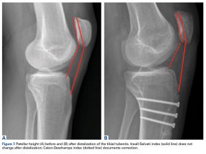
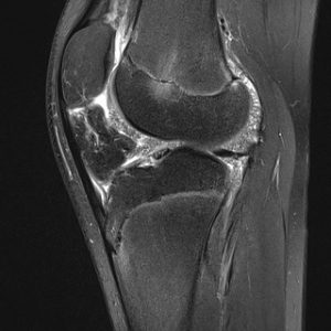
The patient had had some X-rays, and I reviewed these with him: they showed evidence of moderate OA in the medial compartment of each knee, with fully-blown patellofemoral OA at the front of both knees.

Summary

So, what was the problem with the treatment plan of the previous consultant?

First, knee replacement surgery is a big deal. It’s a really big painful operation with very difficult post-op rehab afterwards, and with significant potential risks. When it goes well, the results can be great, but when there’s a problem, it can be catastrophic.

If a patient is ‘young’ (which is very much just an arbitrary concept), then we try to avoid knee replacement if possible (unless it’s a last resort), because the younger a patient is when they have a knee replacement, the bigger the risk of it failing (wearing out) and ending up needing a re-do (a revision knee replacement) within their time. Revision knee replacement is twice as big/complex as a primary, with double the potential complication risk, with slower recovery, with poorer outcomes and with higher re-revision rates. However, if you perform knee replacement surgery in someone in their 70’s, then the risk of them ending up needing a revision within their lifetime is only about 10%, and in about 90% of patients the prosthesis will end up outlasting them.

With older patients, however, the focus often tends to be more on the potential associated risks of the surgery… The older one is, and also the more medical problems one has, then the lower one’s physiological reserve is, in terms of whether the patient is likely to be able to cope with the actual surgery itself. Also, the older one is and the weaker one is, then the less likely they are to be able to cope with the extensive difficult post-op rehab that’s required after knee replacement surgery – and if you don’t do the proper rehab after a knee replacement, then you’re far less likely to end up with a good outcome (i.e. there’s a higher risk of the patient ending up with a stiff and painful knee, with them ending up very unhappy indeed).

It’s hard enough to do the rehab after single unilateral knee replacement surgery. It’s doubly hard to do sufficient rehab if you have both knees done together, at the same time.

Next, this patient was booked in for major surgery very quickly indeed, with minimal time to think things over, to do his own reading or to ask questions, and certainly with no time to do any prehab.

Personally, I always advise my patients to do as much prehab as they possibly can ahead of any knee replacement surgery. Importantly, the fitter and stronger a patient can get themselves ahead of any knee replacement surgery, then the better they’re likely to cope with the op and with the post-op rehab afterwards. Also, if a patient does really well with their prehab, and if they actually end up doing so well that they’re feeling significantly better… then they might even end up not actually needing / wanting the surgery (or at least not, perhaps, for a while longer).

Next, and very importantly… this patient’s main complaint (if you cared to ask the right questions and actually listen properly to what he had to say) was of muscle pains and stiffness, and in clinic he was repeatedly rubbing and massaging specifically his quads muscles.

OK, the patient’s X-rays showed medial plus patellofemoral OA in both knees; however, when I examined the patient’s knees, both knees had a full and pain-free range of motion. Yes, the patient was complaining of pain on stairs, and he was having to go up and down stairs just one step at a time; however, the patient’s patellar tracking looked central and his Clark’s test for patellofemoral irritability was negative at the front of each knee.

Importantly, one should treat the patient, not just the imaging!

The key potential culprit that jumped out at me when I saw this patient in clinic was that he was on statins!

Statins are well-known (or, at least well-known by anyone who actually cares to read the warning inserts that come with every box of tablets, which list the potential side effects) to cause muscle pains, muscle stiffness and muscle weakness … all of which match the exact complaints of this specific patient.

My proposed plan of action

So, what was my advice to this patient?….

  1. First, I advised the patient to cancel (or at least postpone) the surgery that was booked in for him for the following week.
  2. I e-mailed the patient a copy of his clinic letter, which was 4 sides of A4.
  3. I e-mailed the patient links to some additional specific articles that I wanted him to read:

– knee arthritis:  https://kneereplacements.co.uk/knee-arthritis/
– the conservative management of the symptoms of knee arthritis:  https://kneereplacements.co.uk/non-surgical-treatments/
– knee replacement surgery:  http://kneereplacements.co.uk/knee-replacement-surgery-performed/
– the risks of knee replacement surgery:  https://kneereplacements.co.uk/potentials-risks-possible-complications-knee-replacement-surgery/
– the timing of when to actually go ahead with knee replacement surgery:  http://kneereplacements.co.uk/right-time-go-ahead-knee-replacement-surgery/

  1. I advised the patient to go through all of this information carefully, and then to e-mail me back with whatever potential further questions he might have.
  2. The patient had a treadmill, a rowing machine and an exercise bike at home; however, he hadn’t used these for a very long time. So, I advised the patient to start using his exercise bike for 20 to 30 mins a day, every day, and I also advised him to go back to his physio for some additional, more-detailed exercise advice, and I advised the patient to commit to a course of rehab treatments.
  3. Finally, I advised the patient to stop his statins for a trial period.

I also advised the patient to get himself a copy of this book and to read it carefully:

A Statin-Free Life, by Dr Asseem Malhotra, Consultant Cardiologist

https://www.worldofbooks.com/en-gb/products/statin-free-life-book-aseem-malhotra-9781529354102

I’ve asked the patient to drop me an update in 2 months’ time, to let me know how he is getting on.


Comments from the patient’s physio:

“The patient was initially presented to me by an experienced personal trainer colleague who expressed concerns about her client’s recommendation for bilateral total knee replacement surgery by a Consultant Orthopaedic Knee Surgeon. She sought physiotherapy input to gain a more comprehensive perspective regarding the patient’s suitability for surgery.

The patient is an 86-year-old male with imaging-confirmed bilateral knee osteoarthritis, currently under consultant care. Historically, he reported intermittent knee pain, with no recent changes in intensity or frequency. However, he clearly described a significant decline in his physical capabilities, notably struggling with walking and performing sit-to-stand movements from chairs, beds and toilets. He acknowledged never having engaged in regular physical activity, and expressed a dislike for exercise.

Clinical examination revealed a full, pain-free range of motion in both knees, and no tenderness upon joint-line palpation. No swelling, effusion, or ligamentous instability was detected. However, he demonstrated substantial bilateral quadriceps muscle weakness, unable to complete the sit-to-stand test without arm support, performing far below the recommended 8-14 repetitions in 30 seconds for his age group.

My assessment concluded that the patient’s primary issue is significant lower limb muscle weakness and overall physical deconditioning rather than joint pathology alone. Consequently, I recommended against the proposed bilateral knee replacement surgery at this stage, due to his poor physical condition. Instead, I advised a three-month intensive rehabilitation program, aimed at improving muscle strength and functional capacity. This strategy would either eliminate or significantly delay the need for surgical intervention, and, importantly, optimise his physical condition should surgery become necessary later.

In reflecting upon this case, I believe the initial surgical recommendation lacked a comprehensive evaluation of the patient as a whole. Critical factors such as his physical fitness, medical history, and social circumstances were not sufficiently considered. The patient currently supports his wife, who requires daily external care, further complicating his situation. Given his marked sarcopenia, undergoing double knee replacement surgery would likely severely compromise his mobility, potentially rendering him bedridden and significantly diminishing his long-term quality of life. Furthermore, the absence of pre-operative rehabilitation planning exacerbates the risk of poor post-operative outcomes.

Lastly, considering the patient’s advanced age, historical aversion to physical activity and existing responsibilities, he appeared to be a poor candidate for such extensive surgical intervention without significant preparatory measures.

Although the patient had been recommended for surgery by a well-known surgeon, I tried to put that aside and look at this patient’s case objectively and without bias. Because my viewpoint was that the patient was not surgically appropriate, I sought a second surgical opinion and referred the patient across to Mr Ian McDermott, who I knew would look at the scenario properly, in detail, considering all of the variables.”


QUESTIONS

So, questions for you….

  1. Do you think that it’s appropriate / sensible to offer a relatively frail 86-year-old gentleman with a long list of medical problems bilateral simultaneous knee replacement surgery?
  2. Do you think it’s appropriate to book a patient in for very major surgery in such a short timeframe, with them not being given much time at all to think things over or do any prehab?
  3. Do you think it’s appropriate for the patient to have his statins stopped for a trial period?
  4. Would you like to know who this surgeon was and where he’s based?

And finally….

A very big ‘well done’ and ‘thank you’ to the patient’s physio, who was brave enough and confident to stand up for his patient and do the right thing by ‘daring‘ to question the opinions, decisions and actions of one of London’s most ‘famous’ knee surgeons. And if that surgeon doesn’t deserve the title of ‘douchebag’, then perhaps you might like to propose some alternative titles that you think might potentially suit better??…


Douchebag Doctor of the Week: Episode 4 – with Special Guest!!


Well, there’s a LOT to unravel and to ponder on with this one… and I’m honoured to have a very special guest at the end, who has kindly added their personal thoughts on this case and on some of the issues raised. This one’s a bit different, because the ‘Douchebag of the Week’ is actually a physio, not a doctor!

See what you make of it….

Case History

A 62-year-old lady started a Couch-to-5K program back in late 2021. With this, she developed pain in her right knee, and this pain persisted.

In February 2022, the patient went to see a physiotherapist, and she was simply given exercises to do.

In January 2024, the patient went to see a different physio. This physio injected the patient’s knee with hyaluronic acid plus steroid (without any imaging first).

In May 2024, the patient was suffering further pain in her knee. The physio therefore injected the patient’s knee again, this time with just hyaluronic acid.

In August 2024, the patient was again suffering ongoing pain. The patient therefore had her knee injected once again by the physio, again with hyaluronic acid.

In October 2024, the patient went back to the physio, and this time both of her knees were injected with hyaluronic acid.

In November 2024, the patient was still suffering ongoing pain, and she therefore went back to the physio. This time he injected her knee with steroid.

Towards the end of November 2024, the patient ended up coming to see me. At that point, the patient had medial joint line pain, with medial joint line tenderness. She had a fixed flexion deformity of 10o, and her flexion was limited to a maximum of about 110o, limited by pain. There was also a very slight varus in the knee, which was fixed, not correctable. The joint felt stable. A McMurray’s test was negative but a Clark’s test for patellofemoral irritability was positive.

I arranged for the patient to have some imaging of her knee. This imaging showed the following:-

1. There was an extensive degenerate tear of the medial meniscus.
2. There was fully-blown osteoarthritis in the medial compartment, with bone-on-bone contact.
3. There was just minor degeneration in the lateral compartment.
4. There was moderate patellofemoral arthritis at the front of the knee.
5. There were multiple loose bodies in the joint.
6. Finally, there was a small effusion in the knee, and a moderate Baker’s cyst posteriorly.

This patient has now undergone a left total knee replacement under my care, with a Conformis iTotal-CR prosthesis. This surgery was performed in mid-February. The patient has done very well. However, now that her left knee is feeling good, the patient is now more aware of similar pains in her other knee. Imaging has confirmed that the right knee is, similarly, also osteoarthritic, and the patient therefore now wants to go ahead with having her right knee replaced too, as soon as possible.

Questions.

  1. Is it appropriate for a patient to be ‘treated’ within the Independent Healthcare Sector without her first having had appropriate imaging (i.e. without a specific diagnosis first)?
  2. Is it appropriate to inject steroid into a patient’s knee as a form of ‘treatment’?
  3. Is it ever appropriate to inject hyaluronic acid into anyone’s knee (or any joint anywhere)?
  4. Was it appropriate for this patient’s physiotherapist to perform so many injections into her knee, repeatedly, or should she have been referred on much sooner?
  5. Do you think this patient was managed appropriately?

Footnotes:


Special Guest:  Katie Knapton’s comments!

Katie Knapton is a Senior Physiotherapist in West Sussex, and she is the Chair of Physio First. Katie came across this series of case reports and she contacted me via LinkedIn. I’ve shared a couple of specific case reports with Katie, and we’ve had some fairly lengthy discussions about the general implications of some of the specific issues that these cases raise. Katie and I don’t agree on absolutely everything (let’s face it, it would be weird for two people to agree with each other on absolutely everything anyway!)… but we agree on most things, and we certainly agree on putting our patients first and on ensuring that each and every patient is managed properly and only offered appropriate treatments.

Katie has very kindly offered her thoughts on some of the issues raised in this particular episode of ‘Douchbag Doctor of the Week’ — which is particularly important, given that the ‘douchebag‘ is actually a physio this time, not a doctor!

So, I’ll like to thank Katie for getting involved, and for her specific comments:


“I welcome this discussion, though obviously disappointed in the case management presented.

The imaging question deserves clarification – I don’t agree that all knee presentations should be imaged at initial assessment, regardless of healthcare sector. Imaging is not required for most initial knee presentations. A full history and objective assessment should be performed to establish a working clinical diagnosis and decide on an appropriate treatment approach and timelines. This applies whether in NHS or independent practice.

Imaging becomes essential when patients aren’t progressing or when the results will alter the treatment approach. When patients are actually deteriorating – as clearly happened here with the development of a fixed flexion deformity – referring on becomes crucial.

We agree entirely on the core issue: the pattern of repeated ineffective interventions over nearly three years represents a breakdown in sound practice principles. Just doing the same thing because you can is not appropriate and not good practice. Importantly, this shouldn’t be just pointing the finger at injections – all our approaches should be reasoned and clearly communicated with patients.

As our profession evolves and we acquire new skills and treatment modalities, continuous reflection becomes essential. This case reminds us that technical ability to perform an intervention must always be balanced with thorough assessment and evaluation of the individual patient – these fundamentals should remain at the core of all we do.

The value in sharing these cases is to strengthen our collective practice through honest reflection, ultimately improving patient care.”

Katie Knapton, Chair, Physio First.


Douchebag doctor of the week: Episode 3.


Hello campers. Episode 3 of ‘Douchebag doctor of the week’ is here, and this one’s a ‘good’ (bad!) one…

Case history.

A 38-year-old man came to clinic recently complaining of couple of years’ history of gradually worsening right anterior knee pain, with pain on stairs and ladders, and pain with kneeling or squatting; however, the patient was relatively fine walking on the flat, and he was still able to manage any distance (i.e. typical patellofemoral symptoms). The patient was unable to run (although this was because of pre-existing hip problems, from FAI).

On examination, the patient’s knee had a full and pain-free range of motion. The patellar tracking looked good, but the patella was prominent, and it did not seem to engage firmly into the trochlear groove until about 60 degrees of knee flexion. The patella was quite mobile laterally with the knee in extension and the quads relaxed, but the patellar apprehension test (for potential instability) was negative. There was slight crepitus in the patellofemoral joint with knee movements, and a Clark’s test for patellofemoral irritability was mildly positive.

The patient had already been to see a well-known surgeon in one of our two most internationally famous university cities. (From my experience, these two universities tend to churn out individuals whose intellectual arrogance is writing cheques that their ability for independent critical thinking and their EQs just can’t cash – but that’s probably for a different blog!) This surgeon advised the patient that he could have either PRP injections into his knee, or that alternatively, he could have articular cartilage grafting on the back of his patella.

I reviewed the patient’s MRI scan with him. This showed:

– normal medial and lateral compartments, and all the ligaments were intact,
– patellar dysplasia,
– no significant trochlear dysplasia,
– good patellar tracking (with the knee in extension),
– a well-placed tibial tuberosity, with a normal TTTG distance,
– the medial retinaculum was intact and there was no evidence of any traumatic damage to the medial-most edge of the patella (i.e. there was no radiological evidence to suggest that there had ever been any patellar dislocations in the past),
– there was clear evidence of patella alta (with an increased Insall-Salvati ratio and a decreased patellotrochlear index,
– there was just slight increased signal in the articular cartilage on the back of the patella (i.e. early wear and tear, or, to use the somewhat old-fashioned term: ‘chondromalacia patellae’), but with no significant fissures or flaps, and with no actual cartilage loss at all, and certainly with no areas of any full-thickness loss with any actual bare bone exposed.

Image taken from: Patella Alta: A Comprehensive Review
of Current Knowledge. Roland M. Biedert, MD, and Philippe M. Tscholl, MD. The American Journal of Orthopedics® November/December 2017
https://cdn.mdedge.com/files/s3fs-public/Document/October-2017/ajo046060290.pdf

My advice to the patient was that if he felt that his anterior knee pain and his functional restrictions were genuinely bad enough to justify the not-inconsiderable pain, hassle and risks of the surgery, then he could have a tibial tuberosity advancement osteotomy, to correct his patella alta (but without the need for this to have to be combined with an arthroscopy of the knee as well, at the same time).

I reassured the patient that there was certainly no indication whatsoever for any kind of articular cartilage grafting type procedure in his knee, and I also explained to the patient that in my opinion, having PRP injections into his knee would be completely pointless.

I e-mailed the patient a load of information to read about patella alta and about tibial tuberosity advancement osteotomy surgery, and at the time of writing this, the patient is currently mulling things over.

So… main points that stood out to me:

1. The first surgeon that this patient saw didn’t explain to him that he had patella alta.
2. The surgeon offered the patient PRP injections.
3. The surgeon also offered the patient surgery, with articular cartilage grafting (of some kind) to his patella, when the patient doesn’t actually even have any articular cartilage loss on the back of his patella!

Questions for the audience:

1. Should I name the surgeon? (With time, some of you may actually eventually start to appreciate that this question is stated in an ironically rhetorical fashion!) 😉
2. Is this surgeon the biggest ‘Douchebag of the week’ so far?
3. Sadly, I’ve got yet more of these ‘horror story’ cases lined up – so, do you actually find these blogs useful, and would you like to me to continue?

Epilogue for some of my esteemed colleagues

I’ve heard that this ‘Douchebag doctor of the week’ has apparently stirred up some discussion and ruffled some feathers within the ranks of some of my esteemed colleagues here in my sunny hometown of London – if you’re one of these individuals, and if for some reason these stories (every one of which is, sadly, real and absolutely true!) in some way offend you… then A) is it potentially because you maybe recognised some of your own traits and behaviours in some of these stories? or B) do you believe that good doctors should simply keep quiet and that we shouldn’t actually warn people about some of the poor behaviour of some of the bad doctors, and C) if you don’t appreciate these blogs, then why on Earth are you actually bothering to read them? – please, just do us both a favour and please just go away.

Love & Light.

Ian


Douchebag doctor of the week: Episode 2.


So, here we go with Episode 2 of our newly-birthed ‘Douchebag of the week’ series…

This story (below) highlights yet another case of poor-quality shoddy patient management, with (hopefully) a few useful lessons to be learnt.

Case history.

A 59-year-old lady came to clinic complaining of a 2-year history of pain and swelling in both knees.

The patient went to see a local orthopaedic surgeon who sent her for a scan of her left knee, and he advised the patient that she had Grade 4 osteoarthritis. The patient was given an injection of hyaluronic acid into her knee (which, unsurprisingly, did nothing), and the patient was referred for physiotherapy.

The patient’s symptoms persisted. The patient therefore went to see her GP, and unfortunately the GP simply went and injected some i.m. steroid into one of her buttocks.

The patient then went to see a surgeon (who I’ve never heard of before) at an ‘internationally famous clinic’ in Central London. This surgeon arranged for some imaging, with X-rays and MRI scans of both knees. Surprisingly, the follow-up appointment was simply just via a telephone consultation, not face-to-face: the surgeon told the patient that she needed a partial knee replacement in each knee, with both knees being done together, at the same time, and he offered her a slot for this surgery for literally the following week! Thankfully, and very sensibly, the patient decided to get a second opinion, instead, and hence she came to see me.

When I saw the patient myself in clinic, she was complaining of pain and swelling in both knees, with pain when weight-bearing and when walking or exercising, plus she also had pain specifically at the front of her knees with stairs / squatting / kneeling. However, she was still able to walk decent distances and she was not actually having to take any painkillers or anti-inflammatories for her knees at all. Importantly, the patient reported that about 2 years previously, she had suffered an attack of uveitis in one of her eyes (and she was under the care of an ophthalmologist for this), plus the patient had also been getting pain in her elbows, her ankles, her toes and some of her fingers.

When I examined the patient myself, she had a fixed varus deformity in both knees, of over 5 degrees on each side; however, both knees actually had a full range of motion, and there was no actual pain on either side with knee movements or even with forced deep flexion. The knees were not hot, red or swollen, and there was no tenderness at all. The both joints felt stable, and McMurray’s test was negative on both sides; however, a Clark’s test for patellofemoral irritability was positive at the front of each knee.

When I reviewed the patient’s imaging, her MRI scans showed evidence of medial compartmental osteoarthritis in both joints, but with advanced patellofemoral degeneration as well. Importantly, however, when these scans were taken, there was a small to moderate effusion in each knee, with evidence of synovitis and with multiple intra-articular rice bodies present.

MRI scan of a child’s knee (not of this patient’s knee) showing intra-articular rice bodies. [Taken from radiopedia.org: https://radiopaedia.org/cases/rice-bodies-juvenile-idiopathic-arthritis]

I explained to the patient that, unfortunately, she actually had clear evidence of a probable inflammatory arthropathy in both of her knees, and I emphasised to the her that what she actually needed, ahead of anything else, was a formal rheumatological opinion.

I also explained to the patient that even though she does have underlying osteoarthritis (in addition to a probable inflammatory arthropathy), I would not consider her an appropriate candidate for just a medial unicompartmental partial knee replacement in either knee as:-

  • First, she clearly needs a proper diagnosis about her probable rheumatological issues.
  • Next, she will need appropriate medical treatment for this, to get the inflammation to hopefully settle down.
  • Next, her varus deformity in each knee is fixed, plus she also has evidence of symptomatic patellofemoral compartment, and hence even without the presence of an inflammatory arthropathy, I would not consider this lady an appropriate candidate for just a medial unicompartmental partial replacement, and instead, I explained to her that if she were to have surgery under my care, then I would specifically recommend a total knee replacement for each knee.
  • Next, I explained that personally, I am not a fan of bilateral simultaneous knee replacement surgery (given that the post-op rehab for just one knee at a time is so difficult, whilst at the same time being so important, in terms of the patient being able to cope with the rehab and hence achieve a good long-term outcome).
  • Next, and very importantly, partial knee replacement surgery is actually contraindicated in the presence of an inflammatory arthropathy!
  • And finally… this lady’s symptoms from her osteoarthritis are not actually bad enough to justify any kind of arthroplasty type surgery at all at this stage anyway!

What lessons can we learn from this?

In my mind, the mistakes that this surgeon at the ‘internationally famous clinic’ made were:-

  1. First, a post-imaging follow-up appointment, where one should go through the imaging with the patient in detail, and where one should then discuss the diagnosis, the potential treatment options, the pros and cons of those options, the possibility of surgery, the potential risks of any surgery, the likely outcomes of any surgery and the post-op rehab that would be required afterwards… is a terribly important conversation to have with a patient, and this is never something that I, personally, would ever do simply just via a phone call.
  2. Next, it feels really quite inappropriate for this patient to have been offered a slot for major surgery for just 1 week later, after just a telephone consult. Personally, I do not feel that this is appropriate, in terms of giving a patient sufficient time to think things over and ask any additional questions that they might have.
  3. Third, the surgeon at the ‘internationally famous clinic’ in London completely missed all of the fairly obvious red flags in the patient’s history and the fairly blatant synovitis and rice bodies that were clearly visible on the patient’s MRI scan, and hence he missed the very important issue of this patient most likely having an inflammatory arthropathy.

So, questions for the audience….

A) Should I name the surgeon?

B) Should I name the clinic?

C) Would you recommend bilateral simultaneous medial unicompartmental partial knee replacement surgery for a lady with a fixed varus deformity in both knees and with evidence of patellofemoral arthritis, and who is complaining of anterior knee pain?

D) Do you think that telephone consultation was sufficient / appropriate for this lady’s case, and would you consider that ‘gold standard’ management?

E) Would you like me to post a follow-up to this story once the patient has actually seen the rheumatologist? And finally…

F) Why are there are so many utter douchebags within my profession, and would you personally classify this surgeon as a douchebag?


Why are there so many utter douchebags within my profession?


I’m coming across so many cases of poor practice on such a regular basis now that I think the time has finally come to share some of these stories. Sadly, they paint a very poor picture of some of my ‘colleagues’ within my profession. However, I think it’s important that people are aware of what’s actually happening out there, as forewarning people might perhaps help save at least one or two patients from simply accepting what is clearly and blatantly poor practice. This story is just one of many similar shockers, which I’ll share with you in due course…

Case History

A lady comes to see me in clinic for a second opinion. She fell skiing a couple of years ago and felt something ‘go’ in her knee, followed by pain and some (but not a lot of) swelling. Afterwards, her knee felt stable. She had an MRI, and was told that she ‘might’ have had an ACL tear, but the surgeon she saw was not sure. The rest of her knee was apparently OK. The decision was made to managed things conservatively, with rehab. At this stage, her knee still felt stable to her.

A year or so later, the patient then slipped and fell down a few steps, twisting her knee, and this was followed by further pain and swelling in the knee. The patient had another MRI: she was then told by her surgeon that she now had a definite ACL tear, plus medial and lateral meniscal tears. The surgeon recommended surgery, with an ACL reconstruction and meniscal trims.

The patient then went and got herself a second opinion from a well-known very high-volume knee surgeon at a ‘famous’ clinic in London. In clinic, this second surgeon looked at the lady’s MRI report, but not the actual imaging. Shockingly, the patient said that this surgeon did not even actually examine the patient’s knee!!?! The surgeon told the patient that she needed an ACL reconstruction, and that “I’ve done thousands of these: I know what you need”.

The patient then came to see me for what was then a 3rd opinion.

Key details when I saw the patient:

  • c/o mild achy pain at front of knee, in region of tibial tuberosity. No medial or lateral joint line pain. No pain posteriorly.
  • Not confident on knee now on stairs, but no episodes of instability at all.
  • No sudden sharp pains, no painful clicking, no catching, locking or giving way.

Examination:

  • No medial or lateral joint line tenderness.
  • No pain posteriorly with forced deep flexion.
  • McMurray’s test negative.
  • Thessaly’s functional test for meniscal tears negative.
  • Anterior drawer negative.
  • Lachman negative.
  • Pivot shift difficult to assess… but not positive.

Up-to-date MRI:

  • relatively minor partial tear of ACL (not fresh),
  • orientation of PCL = normal (i.e. no anterior tibial subluxation, implying that there is still decent tension in the remnant of the ACL),
  • other ligaments all intact,
  • extensive complex degenerate tear in posterior horn of medial meniscus,
  • complex tear in posterior horn of lateral meniscus as well,
  • no articular cartilage damage and
  • patellofemoral compartment fine.

So… the ‘top’ surgeon that this lady went to see was actually encouraging this patient to go ahead with an ACL reconstruction

  1. when she’s not complaining of any functional instability,
  2. with a negative Lachman’s and a negative pivot shift,
  3. when her MRI scan actually showed that her ACL injury was only a relatively minor partial tear, and, most shockingly…
  4. without him even having examined her knee!!!

My suggested plan of action:

  1. Despite the clear evidence on the MRI of medial and lateral meniscal tears, the patient does not actually seem to be complaining of any specific meniscal type symptoms at present, and her clinical examination failed to show any specific positive meniscal signs. Therefore, there is no need to rush in with an arthroscopy specifically just to deal with the meniscal tears (with what would definitely end up being major meniscal trims, not repairs).
  2. I’ve referred the patient to one of our really excellent local physiotherapists, for some gentle cautious rehab.
  3. The patient has a significantly elevated BMI and expressed concern about this: I’ve therefore also referred the patient to a weight-loss specialist for further advice/assistance.
  4. I’ve sent the patient a load of information to read about:
    • meniscal tears, meniscal repair and meniscal trimming,
    • the consequences of meniscal damage / loss,
    • the role of knee arthroscopy in general,
    • a copy of the latest professional guidelines from The British Association for Surgery of the Knee (BASK) about the appropriate management of meniscal tears and about the role (or otherwise) of knee arthroscopy,
    • ACL tears,
    • ACL reconstruction, and
    • the pros and cons of surgical reconstruction of ACL tears vs conservative management.
  5. I’ve offered to review the patient back in clinic in a couple of months’ time with a fresh up-to-date 3-Tesla MRI scan so that we can then take another look at things and so that we can then chat further… but (hopefully) by then, the patient will know a lot more about her knee issues and the various potential options open to her, and by then she will hopefully be far better informed, and therefore in a much better position to make a proper fully-informed decision about what she might or might not want to have done to her knee.

So… questions for you all:

  1. Do you want me to name the Consultant Orthopaedic Surgeon in London who is a well-known very high-volume knee surgeon at a very well-known clinic?
  2. Should that surgeon be reported for poor practice, for recommending major invasive surgery for a patient A) without looking at their MRI himself (he only looked at the report), B) for not having actually examined the patient’s knee, and C) for advising the patient that she needed to have her knee stabilised when the knee is not actually even unstable?
  3. Do you agree with my slower, more cautious, more measured and more conservative approach
  4. Would you like me to post a follow-up to this sorry story in a couple of months’ time, when the patient (potentially) comes back to clinic again?
  5. Why are there so many utter douchebags within my profession?